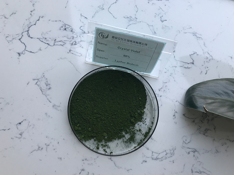
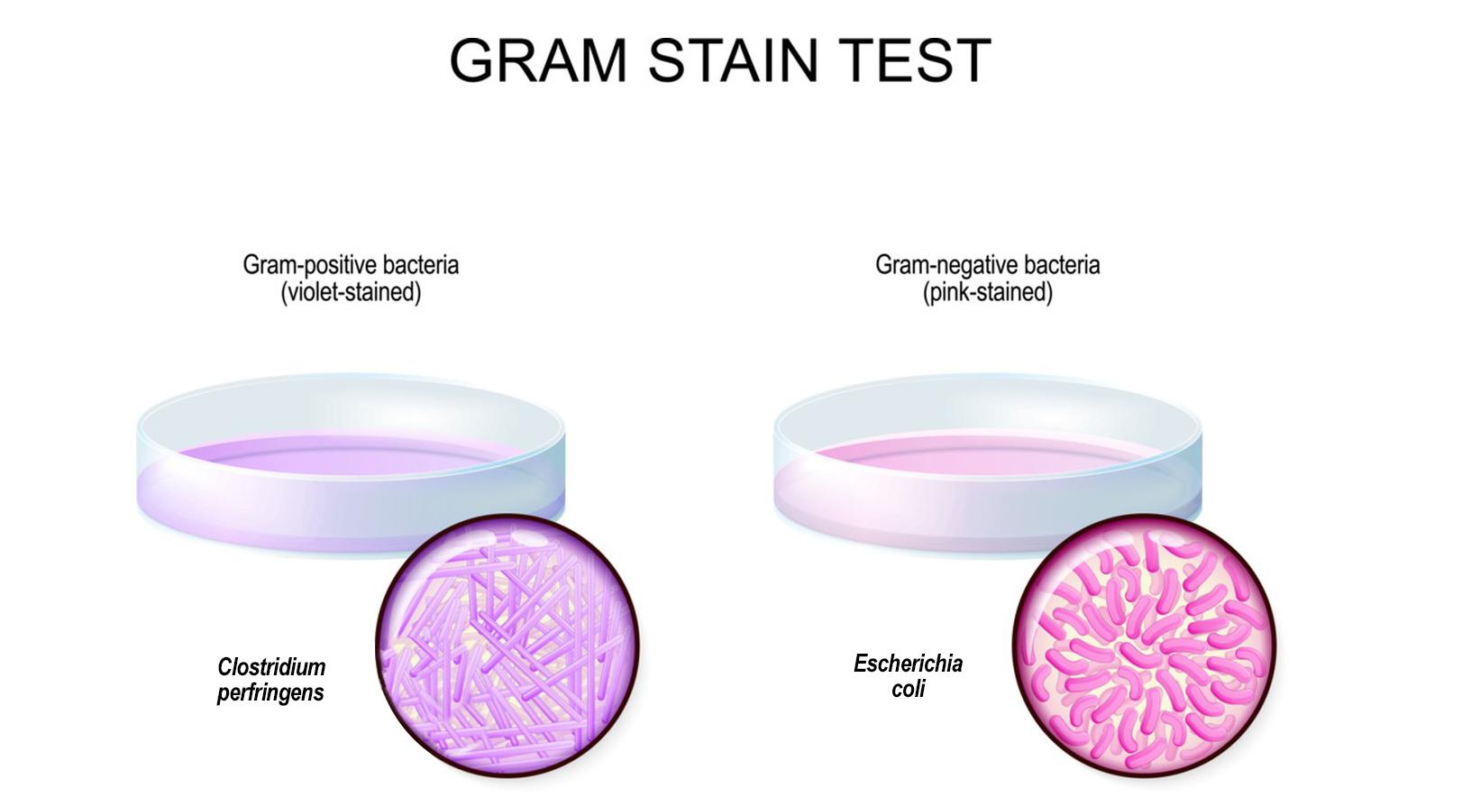
Gentian violet are both synthetic dyes commonly used in laboratory settings, particularly in microbiology. They are often employed as staining agents to visualize and differentiate various biological specimens. Here are the general materials and methods for using Crystal Violet or Gentian Violet in a staining procedure:
Materials for Gentian violet:
1.Crystal Violet or Gentian Violet Solution:
You can purchase pre-made solutions or prepare them by dissolving the dye in an appropriate solvent (usually water or ethanol).
2.Microscope Slides:
Clean glass slides for mounting specimens.
3.Microscope Cover Slips:
Small coverslips placed over the stained specimen.
4.Bunsen Burner or Microscope Slide Warmer:
Used to heat-fix the specimen to the slide.

5.Bacterial Culture or Specimen:
The material you want to stain, such as a bacterial smear on a slide.
6.Staining Tray or Dish:
Container for holding the staining solution.
7.Pipettes:
For dispensing the staining solution onto the specimen.
8.Timer:
To accurately time the staining process.
9.Wash Bottle:
Used for rinsing the specimen during the staining process.
Methods for Gentian violet:
1.Preparation of Specimen:
Prepare a bacterial smear on a clean microscope slide. Allow the smear to air dry.
2.Heat Fixation:
Pass the slide through the flame of a Bunsen burner or use a slide warmer to heat-fix the specimen. This helps to adhere the specimen to the slide.

3.Staining:
Flood the slide with Gentian Violet solution, ensuring that the entire smear is covered. Allow the staining solution to act for a specified period, usually around 1 minute.
4.Rinsing:
Rinse the slide with water or an appropriate rinsing solution to remove excess stain.
5.Drying:
Allow the slide to air dry or use a gentle stream of air to speed up the drying process.
6.Microscopic Examination:
Once the slide is dry, place a coverslip over the stained specimen and examine it under a microscope.
Note: The specific staining time and concentrations may vary depending on the staining protocol you are following or the type of specimen you are working with. Always refer to the specific protocol or method provided by your laboratory or scientific resource.
It’s important to handle these chemicals and materials with care, following appropriate safety guidelines and protocols.
